Bone Cross Section | The superior, the apex of the wedge, is narrow, convex, smooth, and articulates with the lunate. This is a high power photo of a single haversian system. In this section, you'll learn and practise 17 different first aid skills. Adjacent to the hamate on the ulnar side, and slightly above it, is the pisiform bone. In birds and mammals, bone marrow is the primary site of new blood cell production or haematopoiesis.
The hamate bone has six surfaces: This photo shows a cross section through bone. It is composed of hematopoietic cells, marrow adipose tissue, and supportive stromal cells. Each epiphysis meets the diaphysis at the metaphysis, the narrow area that contains the epiphyseal plate (growth plate), a layer of hyaline (transparent) cartilage in a growing. Learn the steps and key action to take when someone needs first aid.

Red marrow fills the spaces in the spongy bone. This is a high power photo of a single haversian system. In this section, you'll learn and practise 17 different first aid skills. Can you identify the primary and secondary haversian systems, central canals and bone lamellae? It is composed of hematopoietic cells, marrow adipose tissue, and supportive stromal cells. In birds and mammals, bone marrow is the primary site of new blood cell production or haematopoiesis. Start with the debate below. This photo shows a cross section through bone. The hamate bone has six surfaces: The films you'll watch cover different first aid scenarios. Learn the steps and key action to take when someone needs first aid. The wider section at each end of the bone is called the epiphysis (plural = epiphyses), which is filled with spongy bone. Each epiphysis meets the diaphysis at the metaphysis, the narrow area that contains the epiphyseal plate (growth plate), a layer of hyaline (transparent) cartilage in a growing.
In this section, you'll learn and practise 17 different first aid skills. Start with the debate below. Red marrow fills the spaces in the spongy bone. The films you'll watch cover different first aid scenarios. Each epiphysis meets the diaphysis at the metaphysis, the narrow area that contains the epiphyseal plate (growth plate), a layer of hyaline (transparent) cartilage in a growing.
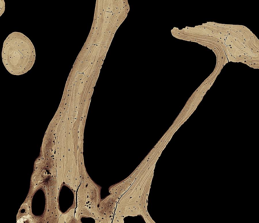
It is composed of hematopoietic cells, marrow adipose tissue, and supportive stromal cells. Adjacent to the hamate on the ulnar side, and slightly above it, is the pisiform bone. Learn the steps and key action to take when someone needs first aid. In this section, you'll learn and practise 17 different first aid skills. The films you'll watch cover different first aid scenarios. This photo shows a cross section through bone. Can you identify the primary and secondary haversian systems, central canals and bone lamellae? This is a high power photo of a single haversian system. Adjacent on the radial side is the capitate, and proximal is the lunate bone.: In birds and mammals, bone marrow is the primary site of new blood cell production or haematopoiesis. Each epiphysis meets the diaphysis at the metaphysis, the narrow area that contains the epiphyseal plate (growth plate), a layer of hyaline (transparent) cartilage in a growing. Red marrow fills the spaces in the spongy bone. Start with the debate below.
The superior, the apex of the wedge, is narrow, convex, smooth, and articulates with the lunate. The films you'll watch cover different first aid scenarios. Each epiphysis meets the diaphysis at the metaphysis, the narrow area that contains the epiphyseal plate (growth plate), a layer of hyaline (transparent) cartilage in a growing. The wider section at each end of the bone is called the epiphysis (plural = epiphyses), which is filled with spongy bone. This photo shows a cross section through bone.
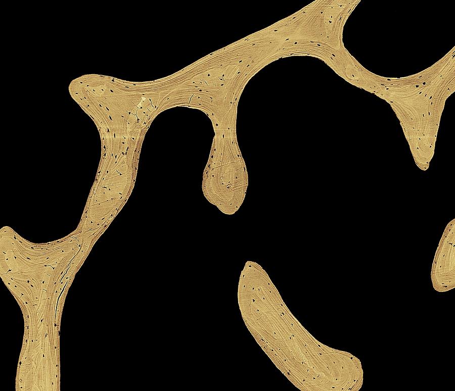
Each epiphysis meets the diaphysis at the metaphysis, the narrow area that contains the epiphyseal plate (growth plate), a layer of hyaline (transparent) cartilage in a growing. Start with the debate below. This is a high power photo of a single haversian system. Adjacent to the hamate on the ulnar side, and slightly above it, is the pisiform bone. The wider section at each end of the bone is called the epiphysis (plural = epiphyses), which is filled with spongy bone. Can you identify the primary and secondary haversian systems, central canals and bone lamellae? The superior, the apex of the wedge, is narrow, convex, smooth, and articulates with the lunate. In this section, you'll learn and practise 17 different first aid skills. Adjacent on the radial side is the capitate, and proximal is the lunate bone.: It is composed of hematopoietic cells, marrow adipose tissue, and supportive stromal cells. Red marrow fills the spaces in the spongy bone. The films you'll watch cover different first aid scenarios. In birds and mammals, bone marrow is the primary site of new blood cell production or haematopoiesis.
Bone Cross Section: In this section, you'll learn and practise 17 different first aid skills.

Tidak ada komentar:
Posting Komentar